Methods For Diagnosing Atrial Fibrillation

The diagnosis of atrial fibrillation is performed primarily through an electrocardiogram. Medical centers have to perform very accurate studies because there are many types of known arrhythmias.
A proper and early diagnosis will allow a patient to cope with the disease better by making changes in their lifestyle and eating habits, as well as establishing a treatment as soon as possible.
Continue reading this article to learn about possible causes and diagnostic methods for this disease.
What is atrial fibrillation?
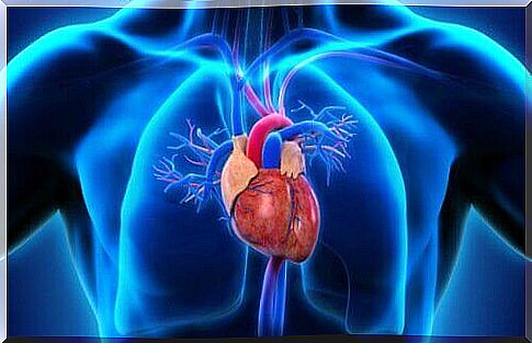
This is a medical term that refers to a heart disease. During atrial fibrillation, the natural electrical signals change. In general, these nerves send impulses that control the contraction and relaxation of the heart. According to the specialized portal Medline Plus, the consequences are as follows:
“Atrial fibrillation can lead to an increased risk of stroke. In many patients, it can also cause chest pain, heart attack or heart failure ”.
What happens is that the atrium (the atrium of the heart) contracts in an irregular and uncoordinated manner in relation to the ventricles (the main chamber of the heart).
The different types of atrial fibrillation and risk groups
This disorder usually occurs in people over 65 years of age. However, there are clinical cases in different age groups. On the other hand, due to unknown causes, it is more common in men than in women.
In the same way, it is possible to distinguish between two types of atrial fibrillation according to their properties :
- Chronic. In this case, the atrial fibrillation has been around for a while and therapy is needed to relieve the symptoms.
- Paroxysmal. This change appears randomly and the associated symptoms correct themselves.
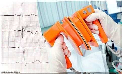
In any case, this disorder can have serious consequences. The most common risks are cerebral infarction and cardiac arrhythmia, or cardiac arrhythmias.
What are the possible causes of atrial fibrillation?
Atrial fibrillation can occur or be triggered by several delayed causes.
To date, researchers have not been able to identify the exact cause or trigger of this disorder. However, there are a number of medical conditions and risks that can lead to atrial fibrillation.
Among them are heart disease or diseases that affect the heart. For example:
- Pericarditis or inflammation of the pericardium (the thin layer that surrounds and protects this organ)
- Myocarditis or inflammation of the myocardium (heart muscle)
- Myocardial infarction
- Valvular heart disease or changes in valves in the heart
- Injuries that have occurred during a surgical operation of the heart
- Smoking and consumption of alcohol and / or drugs
- Drugs that can cause heart damage
- Diseases of the respiratory system such as COPD
- Hyperthyroidism
- Other disorders such as sleep apnea
How Doctors Diagnose Atrial Fibrillation
Doctors will perform a series of medical tests to diagnose atrial fibrillation. In this way, they will be able to rule out other disorders with similar characteristics.
The most common diagnostic methods are :
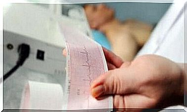
Electrocardiogram or ECG
Doctors perform this test by placing some devices called electrodes on the patient’s chest and arms. These devices are designed to capture the electrical signals that control the movements of the heart. Then a graphical representation of these nerve impulses is displayed.
It is one of the most important diagnostic tests for atrial fibrillation. It can also be found in the form of:
- Holter monitoring. This is a portable ECG device where the patient records the activity of the heart for 24 hours or more.
- Monitor to detect occasional events. In this case, the patient activates the device when they have symptoms of arrhythmia. In this way, a patient can easily get a study of the change timing. Unlike the Holter monitor, the study is performed over a period of several weeks or months.
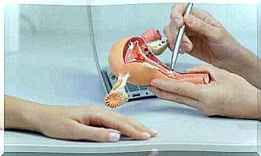
Echocardiogram
Specialists project a set of waves through a device called a transducer to the patient’s chest. The waves hit the heart and bounce back to escape out of the chest cavity. Later, after computer treatment, a live image of the patient’s heart is displayed.
This procedure is called a transthoracic echocardiogram. However, physicians can also insert it through the patient’s mouth using a transducer attached to a flexible tube. When it reaches the esophagus, the test is complete and more detailed details are obtained.
In this way, specialists can check the structure of the organ and see if there are any blood clots. This is a crucial test for diagnosing atrial fibrillation.
Other methods
- Achieve inner images. As a general rule, an x-ray can check the condition of the lungs and heart.
- Stress test or exercise. A patient performs a short physical activity while a team of specialists assesses the heart’s response.
- Routine tests. Doctors usually require a blood test to rule out any underlying disease. They can do this in hyperthyroidism (high levels of thyroid hormones in the blood), for example. You can also perform other tests to see if the patient has a respiratory disorder (usually COPD).