Diagnosis And Treatment Of Pericardial Effusion
Pericardial effusion is a condition that involves fluid retention around the heart. Sometimes it is related to a disease. Other times, however, specific monitoring and evaluation are needed to determine the cause. In some cases, it may be impossible to identify a cause. In fact, it can even become chronic without causing serious problems. However, it is very important in the treatment of pericardial effusion.
Usually, the treatment focuses on solving the problem that is causing the pericardial effusion and on managing the symptoms. If the cause is unknown, the clinical treatment is the same as for pericarditis.
What is pericardial effusion?
Pericardial effusion is an abnormal accumulation of fluid in the pericardial cavity. The pericardium is made up of two layers: a serous layer and a fibrous layer. The distance between these two layers is the heart sac. It normally contains up to 50 ml of serous fluid. When there is an inflamed or infectious process, fluid production increases and pericardial effusion occurs.
This can also manifest itself due to a reduced reabsorption of the liquid. In general, this occurs due to increased systemic venous pressure. In turn, the increase in pressure usually occurs due to congestive heart failure or pulmonary hypertension.
Diagnosis
The clinical presentation of pericardial effusion depends on the rate at which fluid accumulates. Typical symptoms are difficulty breathing and chest pain. Nausea, dysphagia, hoarseness and hiccups are also common symptoms.
When a medical professional suspects a case of pericardial effusion, they may request one or more of the following tests:
- Echocardiogram. This allows the doctor to detect the size of the effusion and evaluate heart function.
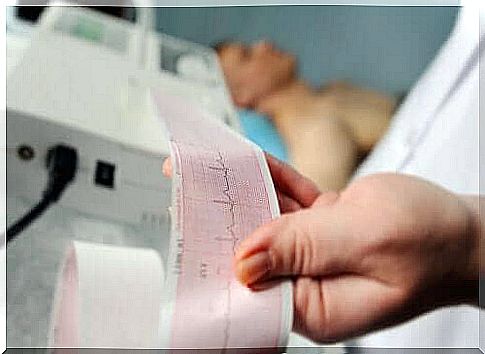
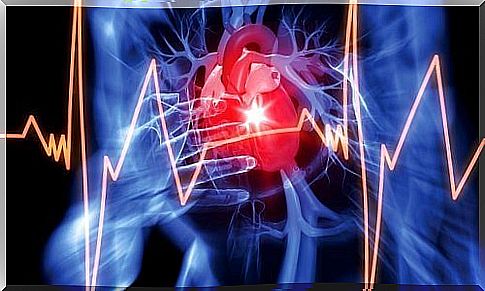
- Electrocardiogram. This test can detect potential blocking patterns.
- X-rays of the chest. These make it possible for medical professionals to determine the size of the pericardial effusion.
The most commonly used diagnostic test is echocardiogram. However, computed tomography (CT) and magnetic resonance imaging (MRI) offer a broader field of view. However, due to availability and cost, medical professionals do not use it as often.
In any case, echocardiographic assessment allows for five important variables: size, duration, distribution, composition, and hemodynamic effects. They used these to determine the cause of pericardial effusion to determine the appropriate course of treatment.
Treatment of pericardial effusion

Overall, treatment of pericardial effusion depends on several factors. These include the amount of fluid accumulated, the existence of cardiac tamponade and the cause. In general, the problem goes away as soon as doctors can treat the cause.
The first step in managing pericardial effusion is to assess its size. Physicians must also define the hemodynamic significance and establish possible associated diseases. It is an underlying disease in about 60% of cases. If there is no tamponade or significant risk of it occurring, most doctors indicate bed rest and anti-inflammatory treatment. Often they also prescribe colchicine and corticosteroids.
However, if there is a risk of tamponade or a high risk of developing the effusion, doctors should perform a pericardiocentesis. When it is not possible to perform this procedure or it fails, open surgical drainage is the next step. This should include a biopsy and the creation of a pericardial window.
Prognosis and monitoring of pericardial effusion
In general, idiopathic pericardial effusion and pericarditis have a good prognosis. Thus, the risk of complications is very low. Cases of chronic idiopathic pericardial effusion have a probability of between 30% and 35% of the scaling to cardiac tamponade. In the second type of effusion, the prognosis depends primarily on the cause that produces it and its size. In about a third of the total cases, those larger than 10 mm worsen and develop into tamponade.
However, it is recommended to monitor moderate idiopathic effusions with an echocardiogram every six months. If severe, the patient should be monitored every three months. In the case of non-idiopathic effusions, monitoring will depend on the disease that caused the pericardial effusion.